Last time I’ve created a set of feline anatomy diagrams, now it’s time for another popular animal—the horse. If you compare them, you’ll notice that thee animals are more similar than they seem at first glance!
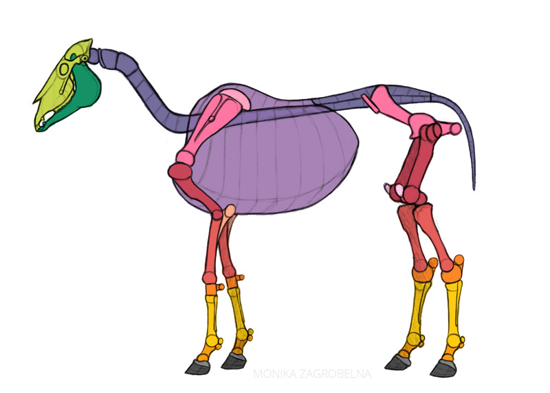
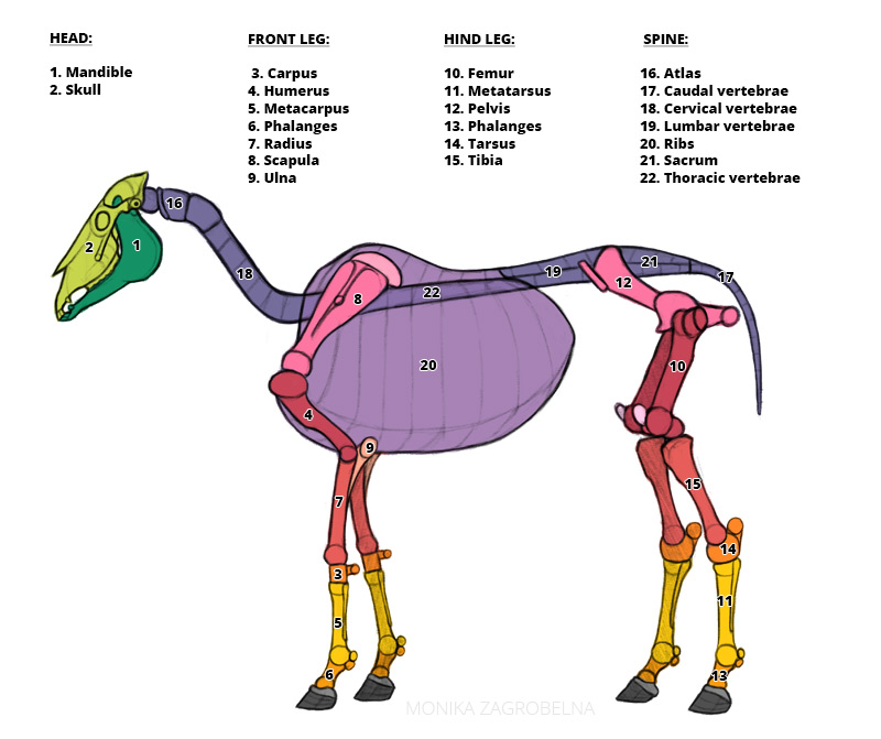
Skeleton
Although bones are mostly hidden from view, understanding their location and shape will help you understand the location and shape of the muscles attached to them. You don’t have to memorize their Latin names—just make sure you know how these bones relate to your own.
Muscles, Layer 1
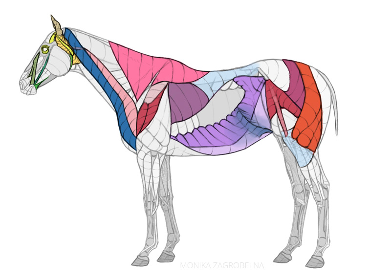
Muscles often overlap each other, so let’s divide them into two separate layers, to analyze them individually. Some of the muscles pictured here are completely covered, but soon you’ll see that they actually affect the outer form of the body.
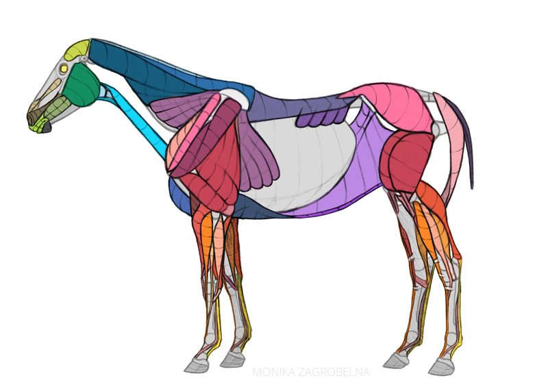
Don’t let these names intimidate you—you don’t have to remember them, but they will be useful if you ever want to learn more abut the specific muscle.
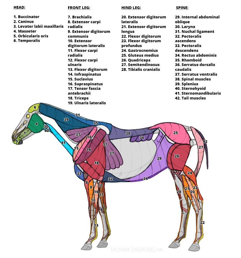
Muscles, Layer 2
These muscles lie more on top, covering the ones from the previous layer.
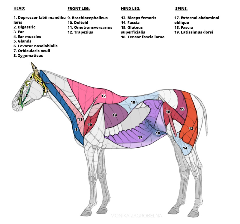
All Muscles + Virtual Dissection
Now you can take a look at how all the muscles look together. I’ve also added the veins to this diagram—they’re often visible on the surface and can be easily confused with an edge of a muscle.
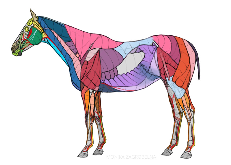
Here you can perform a virtual dissection of a horse! Notice how the form of the muscles affects the final form of the body, and how shading accentuates it.
Can you see how the deeper muscles are visible on the surface? That’s why it’s so important to pay attention to both layers of muscles—if you only use one, complete diagram, you won’t be able to create this effect.
If You’re seriously interested in animal anatomy, here you can find the resources I’ve been using in my studies:
Also, you may also be interested in my animal anatomy course—over 4 hours of narrated video, with exercises, references sheets, and feedback from me!
If you have any questions about these diagrams, or animal anatomy in general, please leave a comment—I’ll be happy to help!

5 Comments